
400-998-5282
专注多肽 服务科研
400-998-5282
专注多肽 服务科研
Kyotorphin是一种内源性神经活性二肽,具有镇痛、神经保护和神经调节特性。Kyotorphin具有抗炎和抗菌活性。它是在神经末梢合成的,存在于大脑和脑脊液中。持续疼痛和阿尔茨海默病患者的脑脊液水平较低。
编号:193905
CAS号:70904-56-2
单字母:H2N-YR-OH
| 编号: | 193905 |
| 中文名称: | 京都肽 Kyotorphin |
| 英文名: | Kyotorphin |
| CAS号: | 70904-56-2 |
| 单字母: | H2N-YR-OH |
| 三字母: | H2N N端氨基:N-terminal amino group。在肽或多肽链中含有游离a-氨基的氨基酸一端。在表示氨基酸序列时,通常将N端放在肽链的左边。 -TyrL-酪氨酸:tyrosine。系统命名为(2S)-氨基-3-(4-羟基苯基)丙酸。是编码氨基酸。符号:Y,Tyr。 -ArgL-精氨酸:arginine。系统命名为(2S)-氨基-5-胍基戊酸。在生理条件下带正电荷,为编码氨基酸。是幼小哺乳动物的必需氨基酸。符号:R,Arg。 -OHC端羧基:C-terminal carboxyl group。在肽或多肽链中含有游离羧基的氨基酸一端。在表示氨基酸序列时,通常将C端放在肽链的右边。 |
| 氨基酸个数: | 2 |
| 分子式: | C15H23N5O4 |
| 平均分子量: | 337.37 |
| 精确分子量: | 337.17 |
| 等电点(PI): | - |
| pH=7.0时的净电荷数: | 2.97 |
| 酸性基团个数: | 1 |
| 碱性基团个数: | 非常亲水 |
| 平均亲水性: | -0.2 |
| 疏水性值: | -2.7 |
| 外观与性状: | 白色粉末状固体 |
| 闪点: | 1280 M-1cm-1 |
| 消光系数: | 1490 |
| 来源: | 人工化学合成,仅限科学研究使用,不得用于人体。 |
| 纯度: | 95%、98% |
| 盐体系: | 可选TFA、HAc、HCl或其它 |
| 储存条件: | 负80℃至负20℃ |
| 标签: | 京都肽(Kyotorphin) 抗菌肽(Antimicrobial Peptides AMPs) 600+种二肽(Dipeptide)现货 |
Kyotorphin是一种内源性神经活性二肽,具有镇痛、神经保护和神经调节特性。Kyotorphin具有抗炎和抗菌活性。它是在神经末梢合成的,存在于大脑和脑脊液中。持续疼痛和阿尔茨海默病患者的脑脊液水平较低。
Kyotorphin is an endogenous neuroactive dipeptide with analgesic, neuroprotective and neuroregulatory properties. Kyotorphin has anti-inflammatory and antibacterial activities. It is synthesized at nerve terminals and found in the brain and cerebrospinal fluid (CSF). People with persistent pain and alzheimer's disease have lower levels of cerebrospinal fluid.
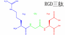
Kyotorphin 是由酪氨酸和精氨酸组成的二肽神经活性分子,可调节神经传递。其结构允许与膜转运体和特定结合位点相互作用。研究人员用它探测实验系统中涉及内源性镇痛通路的信号传导途径。该肽有助于非传统神经肽信号传导的研究。
Kyotorphin is a dipeptide neuroactive molecule composed of tyrosine and arginine that modulates neurotransmission. Its structure allows interaction with membrane transporters and specific binding sites. Researchers use it to probe signaling pathways involving endogenous analgesic pathways in experimental systems. The peptide aids studies of unconventional neuropeptide signaling.
Definition
Kyotorphins are endogenous peptides that take part in the regulation of various adaptive reactions of the organism. They play a role in pain modulation in the mammalian CNS (central nervous system).
Discovery
Kyotorphins (Tyr-Arg) is a dipeptide originally found in bovine and rat brain synaptosomes, is formed from tyrosine and arginine by a specific synthetase. Kyotorphins is a neuroactive peptide named after its place of discovery, Kyoto, Japan1. It has a specific receptor coupled to G i and phospholipase C and elicits enkephalin release.
Structural Characteristics
At biological pH kyotorphins have a neutral net charge. The phenolic rings interact with phospholipid molecules (partition coefficient varies from 6 × 10² to 2 × 10⁴, depending on the lipid and pH used) despite being exposed to the aqueous bulk medium. The lowest energy transition dipole moment is displaced from the normal to the lipid bilayer by 20° on average. The observed extensive interaction, pKa, precise location, and well-defined orientation in membranes combined with the ability to discriminate rigid raft like membrane domains suggest that kyotorphin meets the structural constraints needed for receptor-ligand interaction. The acylated kyotorphin derivative mimics kyotorphin properties and represents a promising way for entrapment in a drug carrier and transport across the blood-brain barrier 2.
Mode of Action
Previous studies suggested that kyotorphin-induced opioid like analgesia may be mediated via a release of Met-enkephalin from the brain. Kyotorphin elicited a release of Met-enkephalin from brain slices but not of [3H]-noradrenaline, [3H]-GABA, [3H]-aspartate and endorphin. The neurochemical basis of mechanisms suggests that Kyotorphins stimulates its specific receptor, followed by G i and phospholipase C (PLC) activations. PLC mechanism leads to a Ca2+ influx in nerve ending particles or synaptosomes. Inositol 1, 4, 5-trisphosphate (InsP3) elicits Ca2+ transport through plasmalemmal InsP3 receptor but not through intra synaptosomal Ca2+ stores. Kyo-induced antinociceptive responses are mediated through its specific receptor. However, at extremely low doses (below femtomolar ranges) of nociceptin/orphanin the endogenous ligand of opioid receptor-like orphan receptor it is coupled to G i, elicits nociceptive responses through its receptor and G i. Potent peripheral nociceptive action of Kyo occur through an InsP3-receptor-gated Ca2+ influx 3,4.
Functions
Kyotorphin improve cardiovascular and cerebral resuscitation after heart arrest - The rate of post resuscitational restoration and survival after a 12-min heart arrest shows that kyotorphin accelerates restoration of vital functions, improve cardiovascular and neurological status within several days after resuscitation 5.
Kyotorphin synthetase activity in rat adrenal glands and spinal cord- Kyotorphin is formed by kyotorphin synthetase from its constituent amino acids, L-Tyr and L-Arg, in the brain in an ATP-Mg2+-dependent manner. To elucidate the physiological role of kyotorphin in organs other than the brain, Kawabata et al., have examined the activity of kyotorphin synthetase in the rat adrenal glands and spinal cord. The activity of adrenal kyotorphin synthetase was inhibited by some L-Arg analogues. Activity was inhibited by NG-nitro-L-arginine methyl ester, alpha-methyl-L-ornithine and D-Arg, but not by NG-nitro-L-arginine and N-iminoethyl-L-ornithine. In the crude soluble extracts from the adrenal glands and spinal cord, kyotorphin was formed by kyotorphin synthetase, and also by the enzymatic processing of the precursor proteins, in the presence of physiological concentrations of L-Tyr and L-Arg in addition to ATP and MgCl2. Kyotorphin synthetase resembling that in the brain is also found to present in the rat adrenal glands and spinal cord, helps in the formation of kyotorphin6.
Kyotorphin suppresses proliferation and Ca2+ signaling in brown preadipocytes-Kyotorphin abolished the stimulatory effect of norepinephrine on proliferation of cultured cells and cold-induced [3H]-thymidine incorporation into DNA of mouse brown adipose tissue in vivo. These changes correlated with peptide-induced suppression of slow calcium signalling in brown preadipocytes.
References
1. Takagi H, Shiomi H, Ueda H, Amano H (1979). A novel analgesic dipeptide from bovine brain is a possible Met-enkephalin release. Nature, 282(5737):410–412.
2. Lopes SC , Soares CM, Baptista AM, Goormaghtigh E, Cabral B , Castanho MA (2006). Conformational and Orientational Guidance of the Analgesic Dipeptide Kyotorphin Induced by Lipidic Membranes: Putative Correlation toward Receptor Docking. J Phys Chem., 110(7):3385–3394.
3. Cheng ZJ, Fan GH, Zhao J, Zhang Z, Wu YL, Jiang LZ, Zhu Y, Pei G, Ma L (1997). Endogenous opioid receptor-like receptor in human neuroblastoma SK-N-SH cells: Activation of inhibitory G protein and homologous desensitization. Neuroreport., 27:1913-1918.
4. Inoue M, Kobayashi M, Kozaki S, Zimmer A, Ueda H (1998). Nociceptin/orphanin FQ-induced nociceptive responses through substance P release from peripheral nerve endings in mice. PNAS, 95:10949-10953.
5. Kharchenko IB, Ziganshin RK, Volkov AV, Koshelev VB (1997). Neokyotorphin and kyotorphin improve cardiovascular and cerebral resuscitation after heart arrest. Bulletin of Experimental Biology and Medicine, 123:450-452.
6. Kawabata A, Muguruma H, Tanaka M, Takagi H (1996). Kyotorphin synthetase activity in rat adrenal glands and spinal cord. Peptide, 17:407-411.
抗菌肽介绍一
AMPs是由相对较小的分子组成的异质基团,通常含有不到100个氨基酸。 它们最初是在20世纪60年代由Zeya和Spitznagel 在多形核白细胞溶酶体中描述的。 迄今为止,已在数据库(如数据库)中 确定和登记了2600多个AMP。 它们是由几乎所有的生物群产生的,包括细菌、真菌、植物和动物。 许多脊椎动物AMPs是由上皮表面分泌的,如 哺乳动物的气管、舌、肠粘膜或两栖动物的皮肤。 有些在中性粒细胞、单核 细胞和巨噬细胞中表达。 AMPs参与动物和植物的免疫防御系统。 构成表达或诱导它们在抵御微生物入侵者 的第一道防线中起着关键作用。
结构/分类 AMPs可以根据其氨基酸组成和结构进行分类。 可以区分两大类AMP。
第一类由线性分子组成,它们要么倾向于采用α螺旋结构,要么富含精氨酸、甘氨 酸、组氨酸、脯氨酸和色氨酸等某些氨 基酸。
第二类由含半胱氨酸的肽组成, 可分为单一或多个二硫结构。 在许多情 况下,抗菌活性需要存在二硫桥。 大多数AMPs是阳离子肽,但也有阴离子肽,如真皮素,一种富含天冬氨酸 的人肽和两栖动物的最大蛋白H5皮肤。 其他非阳离子AMPs包括神经肽前体分子的片段,如原啡肽A, 芳香二肽主要从二翅目幼虫中分离出来,或从节肢动物或茴香物种的氧结合 蛋白中提取的肽。
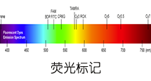
专肽生物可定制合成各类序列的抗菌肽,可标记FITC/FAM/TAMRA等常见荧光素。
Definition
Antimicrobial peptides (AMPs) are as widespread as bacterial inactivator molecules in the innate immune systems of insects, fungi, plants, and mammals. These peptides are also known as host defense peptides (HDPs) as they have other immuno-modulatory functions besides the direct antimicrobial actions and are even capable of killing cancerous cells 1,2.
Classification
Three broad categories of HDPs have been identified: 1) the linear peptides with helical structures, 2) the cysteine stabilized peptides with beta-sheet, and 3) a group of linear peptides rich in proline and arginine that primarily have been identified in non-mammalian species.
Structural characteristics
In mammals, cathelicidins and defensins are the two principal AMP families. Cathelicidins are peptides with a conserved proregion and a variable C-terminal antimicrobial domain. Defensins are the best-characterized AMPs, they have six invariant cysteines, forming three intramolecular cystine-disulfide bonds.
Mode of action
The mode of action of AMPs elucidated to date include inhibition of cell wall formation, formation of pores in the cell membrane resulting in the disruption of membrane potential with eventual lysis of the cell. These peptides also inhibit nuclease activity of both RNase and DNase.
Functions
They have a broad ability to kill microbes. AMPs form an important means of host defense in eukaryotes. Large AMPs (>100 amino acids), are often lytic, nutrient-binding proteins or specifically target microbial macromolecules. Small AMPs act by disrupting the structure of microbial cell membranes. It plays an active role in wound repair and regulation of the adaptive immune system. They have multiple roles as mediators of inflammation with impact on epithelial and inflammatory cells, influencing diverse processes such as cell proliferation, wound healing, cytokine release, chemotaxis and immune induction 3.
References
1. Gottlieb CT, Thomsen LE, Ingmer H, Mygind PH, Kristensen HH, Gram L(2008). Antimicrobial peptides effectively kill a broad spectrum of Listeria monocytogenes and Staphylococcus aureus strains independently of origin, sub-type, or virulence factor expression. BMC Microbiol., 8:205.
2. Yeaman MR and Yount NY (2003). Mechanisms of Antimicrobial Peptide Action and Resistance. Pharmocological Reviews, 55(1).
3. Hanna Galkowska H and Olszewski WL (2003). Antimicrobial peptides – their role in immunity and therapeutic potential. Centr Eur J Immunol., 28 (3):138–141.
抗菌肽介绍二
Ribosomally synthesized antimicrobial peptides (AMPs) constitute a structurally diverse group of molecules found virtually in all organisms. Most antimicrobial peptides contain less than 100 amino acid residues, have a net positive charge, and are membrane active. They are major players in the innate immune defense but can also have roles in processes as chemokine induction, chemotaxis, inflammation, and wound healing. In addition to their antimicrobial effects, many of them show antiviral and antineoplastic activities.
INTRODUCTION
AMPs are a heterogeneous group of relatively small molecules usually containing less than a hundred amino acids. They were first described in the 1960’s by Zeya and Spitznagel in polymorphonuclear leukocyte lysosomes.
To date, more than 2600 AMPs have been identified and registered in databases. They are produced by nearly all groups of organisms, including bacteria, fungi, plants, and animals. Many vertebrate AMPs are secreted by epithelial surfaces such as the tracheal, lingual, or intestinal mucosa of mammals or the skin of amphibia. Some are expressed in neutrophils, monocytes, and macrophages.
AMPs are involved in both animal and plant immune defense systems. Constitutively expressed or induced they play a key role in the first line of defense against microbial intruders.
STRUCTURE/CLASSIFICATION
AMPs can be classified on the basis of their amino acid composition and structure. Two major groups of AMPs can be distinguished. The first group consists of linear molecules which either tend to adopt α-helical structure or are enriched in certain amino acids such as arginine, glycine, histidine, proline, and tryptophan. The second group consists of cysteine-containing peptides which can be divided into single or multiple disulfide structures. In many cases, the presence of disulfide bridges is required for antimicrobial activity.
Most AMPs are cationic peptides, but there are also anionic peptides such as dermcidin, an aspartic acid-rich peptide from human and maximin H5 from amphibian skin. Other non-cationic AMPs include fragments from neuropeptide precursor molecules such as proenkephalin A, aromatic dipeptides primarily isolated from dipteran larvae, or peptides derived from oxygen-binding proteins from arthropod or annelid species.
MODE OF ACTION
Most AMPs act by provoking an increase in plasma membrane permeability. They preferentially target microbial versus mammalian cells. Selectivity is influenced by several factors such as differences in membrane composition: membranes of many bacterial pathogens contain negatively charged lipid moieties such as phosphatidylglycerol (PG), cardiolipin, and phosphatidylserine (PS), whereas mammalian membranes, commonly enriched in phosphatidylethanolamine (PE), phosphatidylcholine (PC) and sphingomyelin, are generally neutral in net charge.
The presence of sterols such as cholesterol and ergesterol within the membrane may be a further means by which AMPs can distinguish between mammalian or fungal cells and prokaryotes. A first step in the mechanism of membrane permeabilization is the electrostatic interaction between the positively charged AMP with the negatively charged membrane surface of the microorganism. Subsequent disruption of the membrane by creation of pores within the microbial membrane ultimately results in cell death of the organism due to leakage of ions, metabolites, cessation of membrane-coupled respiration, and biosynthesis.
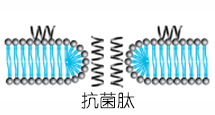
Several models for pore formation such as the Barrel-Stave, the Toroidal or Wormhole Model, and the Carpet Model have been proposed (Fig. 1).
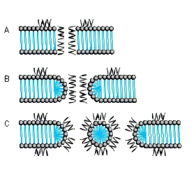
FIG. 1. MODE OF ACTION A BARREL-STAVE MODEL B TOROIDAL PORE OR WORMHOLE MODEL C CARPET MODEL
THE BARREL-STAVE MODEL
The Barrel-Stave model describes a mechanism in which AMPs form a barrellike pore within the bacterial membrane with the individual AMPs or AMP complexes being the staves. Arranged in this manner, the hydrophobic regions of the AMPs point outwards towards the acyl chains of the membrane whereas the hydrophilic areas form the pore.
THE TOROIDAL PORE OR WORMHOLE MODEL
The pores described by this model differ from those of the Barrel-Stave model. Primarily, the outer and inner leaflet of the membrane are not intercalated in the transmembrane channel.
THE CARPET MODEL
A different mechanism is proposed in the Carpet model where AMPs first cover the outer surface of the membrane and then disrupt the membrane like detergents by forming micelle-like units. Certain AMPs penetrate the bacterial membrane without channel formation. They act on intracellular targets by e.g. inhibiting nucleic acid and/or protein synthesis.
RESISTANCE
Resistance to AMPs can either be constitutive or inducible. Inherited resistance mechanisms include altered surface charge, active efflux, production of peptidases or trapping proteins, and modification of host cellular processes. For instance, Staphylococcus aureus manages to reduce the overall cell surface charge by esterification of the cell wall component teichoic acid with D-alanine and thereby increases its resistance against human AMPs. Another example for changing the surface net charge is the production of cationic lysine-substituted phosphatidylglycerol (L-PG) found in certain Staphylococcus aureus strains. In Gram-negative bacteria, addition of 4-aminoarabinose (Ara4N) to the phosphate group of the lipid A backbone or increased acylation of lipopolysaccharides (LPS) are important mechanisms of AMP resistance. Exposure to AMPs may also induce stress responses by which microorganisms try to survive. Inducible regulatory mechanisms have been described in a variety of organisms. For instance, the PhoP/PhoQ regulon in Salmonella has been demonstrated to regulate transcriptional activation of surface and secretory proteins, enzymes that modify lipopolysaccharide, lipid and protein constituents of the outer membrane and proteases that likely degrade certain AMPs.
EXAMPLES OF ANTIMICROBIAL PEPTIDES
| Cationic peptides enriched for specific amino acids |
|
|---|---|
| Glycine-containing peptides | Hymenoptaecin from honeybees |
| Glycine- and proline-containing peptides | Coleoptericin from beetles Holotricin from beetles |
| Histidine-containing peptides | Histatins from humans and some higher primates |
| Proline-containing peptides | Abaecin from honeybees |
| Proline- and arginine-containing peptides | Apidaecins from honeybees Bactenicins from cattle Drosocin from Drosophila PR-39 from pigs |
| Proline- and phenylalanine-containing peptides | Prophenin from pigs |
| Tryptophan-containing peptides | Indolicidin from cattle |
| Linear cationic α-helical peptides | |
|---|---|
| Andropin from insects Bombinin from amphibians Buforin II from amphibians CAP18 from rabbits Cepropins from insects Cecropin P1 from the pig intestinal parasitic nematode, Ascaris suum Ceratotoxin from insects Dermaseptin from amphibians LL-37 from human Magainin from amphibians Melittin from insects Pleurocidin from Pseudopleuronectes americanus |
| Anionic and cationic peptides that contain cysteine and form disulfide bonds |
|
|---|---|
| 1 Disulfide bond | Brevinins |
| 2 Disulfide bonds | Protegrins from pigs |
| 3 Disulfide bonds | α-Defensins from human, rabbits and rats β-Defensins from humans, cattle, mice, rats, pigs, goats and poultry θ-Defensin from the rhesus monkey Insect defensins (Defensin-A from Aedes aegypti) |
| 4 Disulfide bonds | Antifungal defensins from plants Drosomycin from Drosophila |
| Anionic peptides | Dermcidin from human skin Maximin H5 from amphibian skin |
| Anionic and cationic peptide fragments derived from precursor proteins |
Antimicrobial domains from bovine α-lactalbumin, human hemoglobin, lysozyme, and ovalbumin Aromatic dipeptides from dipteran larvae Casocidin I from human casein Enkelytin from proenkaphalin A Lactoferricin from lactoferrin |
ADAPTED FROM K.A. BROGDEN, NAT. REV. MICROBIOL. 3, 238-250 (2005)
IMPORTANT FAMILIES OF AMPS
BOMBININS
Bombinins constitute a family of AMPs produced in fire-bellied toads (Bombina species) active against Gram-negative and Gram-positive bacteria and fungi. Bombinins, bombinin-like peptides (BLPs), and Bombinin H molecules are found in the species Bombina bombina, Bombina variegata, and Bombina orientalis, whereas the homologous maximins and maximin H peptides are derived from the giant fire-bellied toad Bombina maxima. Bombinin H peptides contain either 17 or 20 amino acid residues and are more hydrophobic than bombinins, some of them contain D-alloisoleucine at position 2. They exhibit lower antibacterial activity than bombinins but, in contrast to them, they possess haemolytic activity.
CATHELICIDINS
Members of this family are amphipathic, cationic peptides with a broad-spectrum antimicrobial activity. Cathelicidins typically act by disrupting the integrity of bacterial membranes. They are characterized by an evolutionary conserved N-terminal cathelin- like domain of approximately 99-114 amino acid residues linked to a C-terminal antimicrobial domain of 12-100 residues that can be released upon proteolytic processing. Members of this family include linear peptides amongst them a number of proline-rich AMPs that show different types of proline repeat motifs (Bac5, Bac7, PR-39, prophenins) and the tryptophan-rich indolicidin characterized by three regularly spaced proline residues. The protegrins (PG-1 to PG-5) contain two disulfide bridges and an amidated C-terminus. Cathelicidins have been found in every mammalian species examined. In human, LL-37 (Product 4042456) is the only member of the cathelicidin family. The peptide consists of 37 amino acids and contains two leucine residues at the N-terminus. It is proteolytically cleaved from the 18 kDa precursor protein human cathelicidin antimicrobial protein CAP-18. LL-37 is primarily produced by phagocytic leucocytes and epithelial cells, and is involved in various processes such as direct killing of microorganisms, binding and neutralizing LPS, chemotaxis and chemokine induction, regulation of inflammatory responses, and wound healing. Its production is influenced by several factors such as microbial products, host cytokines, vitamin D3, and availability of oxygen. LL-37 orthologues in mouse and rat are CRAMP (mouse) (Product 4056438) and CRAMP (rat), respectively.
CECROPINS
Cecropins were first isolated from the giant silk moth Hyalophora cecropia. They can form amphipathic, α-helical structures and are structurally related to other cecropins as bactericidin, lepidopteran, and sarcotoxin. Cecropin P1 (Product 4039862), found in pig intestine, also belongs to this family. Most cecropins show broad-spectrum antibacterial activity. Cecropin A (Product 4030488) and B (Product 4030477) have also been demonstrated to possess tumoricidal activity against mammalian leukemia, lymphoma, and carcinoma cell lines.
CERATOTOXINS
This family consists of cationic α-helical amphipathic peptides expressed in the female reproductive accessory glands of the Mediterranean fruit fly Ceratitis capitata. The production of the peptides is enhanced by mating. Ceratotoxin A and ceratotoxin B are 29 amino acid peptides differing in two amino acids. Ceratotoxin C and D consist of 32 and 36 amino acids, respectively. The peptides of this family are active against Gram-negative as well as Grampositive bacteria and are supposed to act via the Barrel-Stave model. Ceratotoxin A has been shown to be mainly antibacterial for Gram-negative organisms.
DEFENSINS
Defensins are small cysteine-rich cationic peptides containing three or four disulfide bridges. They have been isolated from molluscs, acari, arachnids, insects, mammals, and plants. They are further divided into families on the basis of the spatial distribution of their cysteine residues. Three families, the α-, β- and θ-defensins, can be distinguished in mammals. α- and β-defensins are characterized by antiparallel β-sheet structures stabilized by three disulfide bonds. The θ-defensins are found in rhesus monkey and some other non-human primates but not in human, chimpanzee and gorilla. They consist of two nine amino acid peptides derived from different precursor proteins joined by head-to-tail cyclization. Invertebrate and plant defensins contain three or four disulfide bridges, respectively. Insect and mammalian defensins are mainly active against bacteria while most plant defensins possess antifungal activity.
DERMASEPTINS
The peptides of the dermaseptin family are closely related and consist of 28-34 amino acids. They were originally isolated from skin extracts of the South American arboreal frog Phyllomedusa sauvagei and contain a conserved tryptophan residue at position 3. Dermaseptins exhibit broad-spectrum antimicrobial activity against Gram-positive and Gram-negative bacteria.
HISTATINS
Histatins are histidine-rich and mostly cationic peptides found in the saliva of humans and some higher primates. They are active against a broad-spectrum of bacteria and fungi. The antifungal activity of the human salivary peptide histatin-5 has been extensively studied and is supposed to be due to inhibition of mitochondrial respiration and the formation of reactive oxygen species. Histatin-5 has also been shown to inhibit both host-derived and bacterial proteolytc enzymes involved in peridontal diseases. Histatin-8, a peptide from human parotid secretion, has been shown to inhibit hemagglutination activity of Porphyromonas gingivalis 381, a Gram-negative bacterium involved in certain forms of periodontal disease. The peptide may function as a binding domain for the hemagglutinins of Porphyromonas gingivalis during agglutination.
MAGAININS
Magainins constitute a family of linear amphipathic cationic AMPs discovered in the skin of Xenopus laevis. The two closely related members of this family, magainin I (Product 4012844) and magainin II (Product 4013706) differ merely in two positions and are 23 amino acids in length. Magainins exhibit broad-spectrum antimicrobial activity against Gram-negative and Gram-positive bacteria, fungi and protozoa and are also cytotoxic for many murine and human cancer cell lines.
CONCLUSIONS
The structures of AMPs represent a unique source for the targeted exploration of new applications in the therapy of microbial and viral infection, cancer, and sepsis. Modern synthetic methods will allow the relatively cheap and accurate production of lead compounds and peptide candidates. The achievements in peptide library generation, analytical methods as mass spectrometry, and screening and formulation technologies may contribute to solve intrinsic problems associated with the use of AMPs as therapeutic agents such as susceptibility to proteases and host toxicity. Bachem has considerable expertise and long-standing experience in peptide synthesis. With our capacity to upscale the production of simple and modified peptides, we are the partner of choice for the pharmaceutical industries.