
400-998-5282
专注多肽 服务科研
400-998-5282
专注多肽 服务科研

Angiogenin (108-123)的序列是ENGLPVHLDQSIFRRP,它对应的是angiogeninC端区域,Angiogenin (108-123)可以抑制angiogenin的酶促和生物学活性。
编号:154101
CAS号:112173-48-5
单字母:H2N-ENGLPVHLDQSIFRRP-OH
血管生成素(108‑123)捕获一段功能区段,用于研究折叠、催化基序和核糖核酸酶相关相互作用。其疏水残基和极性残基支持结构分析和构象模拟。该多肽有助于定位母蛋白内的结合热点。应用包括酶促机制研究和蛋白质‑多肽识别研究。
Angiogenin (108-123) captures a functional segment used to investigate folding, catalytic motifs, and ribonuclease-related interactions. Its hydrophobic and polar residues support structural analysis and conformational modeling. The peptide aids in mapping binding hotspots within the parent protein. Applications include enzymatic mechanism studies and protein-peptide recognition research.
Angiogenin (108-123)的序列是ENGLPVHLDQSIFRRP,它对应的是angiogeninC端区域,Angiogenin (108-123)可以抑制angiogenin的酶促和生物学活性。
Definition
Angiogenin (ANG), a 14,124 Da protein that has been implicated in angiogenesis in tumor progression. It is secreted by tumor cells, a potent inducer of neovascularization1.
Discovery
In 1885, Fett et al., first isolated and characterized angiogenin from medium conditioned by HT-29 human colon adenocarcinoma cells1 and later from normal mammalian plasma2 and milk. It has been subsequently isolated from human, bovine, rabbit, pig & mouse sera and bovine milk. Angiogenin has ribonucleolytic activity with 33% sequence homology to pancreatic RNAse A3. A synthetic peptide ’H-Glu-Asn-Gly-Leu-Pro-Val-His-Leu-Asp-Gln-Ser-Ile-Phe-Arg-Arg-OH (108-122) corresponding to the C-terminal region of ANG inhibits the enzymatic and biological activities of angiogenin4. Several C-terminal synthetic peptides, including (Ang 108-123), significantly decreases angiogenin-induced neovascularization.
Structural characteristics
Acharya et al., determined the crystal structure of human antigenic at 2.4 0A. Overall Structure features a kidney shaped tertiary fold reminiscent of RNase A. The ribonucleolytic active center (His-13, His-114, and Lys-40) and the putative receptor binding site, both of which are critically involved in biological functions are distinct from Rnase A5. The central core of the molecule consists of P structure with a pair of antiparallel twisted forming the main topology with residues Ser-72 and Gly-99 at the apices. Two additional strands on either side of these central strands (residues 41-47; 111-116) complete the major sheet structure. There are 3 helices H1, residues 3-14, H2, residues 22-33 and H3, residues 49-58 present in the structure.
Mechanism of action
Four aspects of ANG have been discovered that is necessary for the process of ANG-induced angiogenesis,ANG exerts its ribonucleolytic activity-ANG has a very weak 105-106 lower ribonucleolytic activity than that of RNase A. This is because the pyrimidine binding site of ANG is “obstructed” by the glutamine (Gln)117 residue. However, ribonucleolytic activity of ANG is crucial for angiogenesis.ANG stimulates basement membrane degradation-ANG binds to a-actin on endothelial cell surface; ANG-actin complexes dissociate from the cell surface and accelerate tissue type plasminogen activator (tPA)-catalyzed generation of plasmin from plasminogen. ANG-actin complexes promote the degradation of basement membrane and extracellular matrix. This complex allows endothelial cells to penetrate and migrate into the perivascular tissue. Basement membrane degradation is an essential feature of angiogenesis. ANG activates signaling transduction- ANG binds to a 170-kDa receptor located on the endothelial cell surface and elicits second messenger systems. Binding of ANG to cell surface actin results in activation of a cell-associated protease system that promotes cell invasion. ERK1/2, protein kinase B/Akt1 pathways have been proposed to be activated by ANG stimulation. ANG nuclear translocation-Angiogenin undergoes nuclear translocation in endothelial cells via receptor-mediated endocytosis and nuclear localization sequence-assisted nuclear import. A nuclear localization signal (NLS), lies in 31-RRRGL-35 of the protein. Upon nuclear translocation it enhances rRNA transcription.
Functions
Functions of ANG in Angiogenesis-As a key angiogenic factor, ANG interacts with endothelial and smooth muscle cells to induce a wide range of cellular responses including cell migration, invasion, proliferation, and formation of tubular structures. ANG has also been reported to induce the proliferation of cancer cells directly. Recently, ANG gene was identified to be a potential amyotrophic lateral sclerosis (ALS) related gene6. ANG induces tumor growth in various types of human cancers, including breast, cervical, colon, colorectal, endometrial, gastric, liver, kidney, ovarian, pancreatic, prostate, and urothelial cancers, as well as astrocytoma, leukemia, lymphoma, melanoma, osteosarcoma, and Wilms’ tumor 6. ANG may be related with amyotrophic lateral sclerosis- Amyotrophic lateral sclerosis (ALS) is a progressive late onset neurodegenerative disorder affecting upper and lower motoneurons (MNs). Vascular endothelial growth factor was the first angiogenic factor shown to contribute to the pathogenesis of ALS 7.
References
1. Fett JW, Strydom DJ, Lobb RR, Alderman EM, Bethune JL, Riordan JF, Vallee BL (1985). Isolation and characterization of angiogenin, an antigenic protein from human carcinoma cells. Biochemistry, 24: 5480-5486.
2. Shapiro R, Strydom DJ, Olson KA, Vallee BL (1987). Isolation of angiogenin from normal human plasma. Biochemistry, 26: 5141-5146.
3. Strydom DJ, Fett JW, Lobb RR, Alderman EM, Bethune JL, Riordan JF, Vallee BL (1985). Amino acid sequence of human tumor derived angiogenin. Biochemistry, 24(20): 5486–5494.
4. Rybak SM, Auld DS, St Clair DK, Yao QZ, Fett JW (1989). C-terminal angiogenin peptides inhibit the biological and enzymatic activities of angiogenin. Biochem. Biophys. Res. Commun, 162: 535–543.
5. Acharya KR, Shapiro R, Allen SC, Riordan JF, Vallee BL (1994). Crystal structure of human angiogenin reveals the structural basis for its functional divergence from ribonuclease. Proc Natl Acad Sci USA, 91: 2915-2919.
6. Yoshioka N, Wang L, Kishimoto K, Tsuji T, Hu GF (2006). A therapeutic target for prostate cancer based on angiogenin-stimulated angiogenesis and cancer cell proliferation. Proc Natl Acad Sci USA, 103: 14519-14524.
7. Oosthuyse B, Moons L, Storkebaum E, Beck H, Nuyens D, Brusselmans K, Van Dorpe J, Hellings P, Gorselink M, Heymans S, Theilmeier G, Dewerchin M, Laudenbach V, Vermylen P, Raat H, Acker T, Vleminckx V, Van Den Bosch L, Cashman N, Fujisawa H, Drost MR, Sciot R, Bruyninckx F, Hicklin DJ, Ince C, Gressens P, Lupu F, Plate KH, Robberecht W, Herbert JM, Collen D, Carmeliet P(2001). Deletion of the hypoxia-response element in the vascular endothelial growth factor promoter causes motor neuron degeneration. Nat Gene, 28:131-138.
多肽H2N-Glu-Asn-Gly-Leu-Pro-Val-His-Leu-Asp-Gln-Ser-Ile-Phe-Arg-Arg-Pro-COOH的合成步骤:
1、合成CTC树脂:称取1.11g CTC Resin(如初始取代度约为0.51mmol/g)和0.68mmol Fmoc-Pro-OH于反应器中,加入适量DCM溶解氨基酸(需要注意,此时CTC树脂体积会增大好几倍,避免DCM溶液过少),再加入1.7mmol DIPEA(Mw:129.1,d:0.740g/ml),反应2-3小时后,可不抽滤溶液,直接加入1ml的HPLC级甲醇,封端半小时。依次用DMF洗涤2次,甲醇洗涤1次,DCM洗涤一次,甲醇洗涤一次,DCM洗涤一次,DMF洗涤2次(这里使用甲醇和DCM交替洗涤,是为了更好地去除其他溶质,有利于后续反应)。得到 Fmoc-Pro-CTC Resin。结构图如下:

2、脱Fmoc:加3倍树脂体积的20%Pip/DMF溶液,鼓氮气30分钟,然后2倍树脂体积的DMF 洗涤5次。得到 H2N-Pro-CTC Resin 。(此步骤脱除Fmoc基团,茚三酮检测为蓝色,Pip为哌啶)。结构图如下:

3、缩合:取1.7mmol Fmoc-Arg(Pbf)-OH 氨基酸,加入到上述树脂里,加适当DMF溶解氨基酸,再依次加入3.4mmol DIPEA,1.61mmol HBTU。反应30分钟后,取小样洗涤,茚三酮检测为无色。用2倍树脂体积的DMF 洗涤3次树脂。(洗涤树脂,去掉残留溶剂,为下一步反应做准备)。得到Fmoc-Arg(Pbf)-Pro-CTC Resin。氨基酸:DIPEA:HBTU:树脂=3:6:2.85:1(摩尔比)。结构图如下:

4、依次循环步骤二、步骤三,依次得到
H2N-Arg(Pbf)-Pro-CTC Resin
Fmoc-Arg(Pbf)-Arg(Pbf)-Pro-CTC Resin
H2N-Arg(Pbf)-Arg(Pbf)-Pro-CTC Resin
Fmoc-Phe-Arg(Pbf)-Arg(Pbf)-Pro-CTC Resin
H2N-Phe-Arg(Pbf)-Arg(Pbf)-Pro-CTC Resin
Fmoc-Ile-Phe-Arg(Pbf)-Arg(Pbf)-Pro-CTC Resin
H2N-Ile-Phe-Arg(Pbf)-Arg(Pbf)-Pro-CTC Resin
Fmoc-Ser(tBu)-Ile-Phe-Arg(Pbf)-Arg(Pbf)-Pro-CTC Resin
H2N-Ser(tBu)-Ile-Phe-Arg(Pbf)-Arg(Pbf)-Pro-CTC Resin
Fmoc-Gln(Trt)-Ser(tBu)-Ile-Phe-Arg(Pbf)-Arg(Pbf)-Pro-CTC Resin
H2N-Gln(Trt)-Ser(tBu)-Ile-Phe-Arg(Pbf)-Arg(Pbf)-Pro-CTC Resin
Fmoc-Asp(OtBu)-Gln(Trt)-Ser(tBu)-Ile-Phe-Arg(Pbf)-Arg(Pbf)-Pro-CTC Resin
H2N-Asp(OtBu)-Gln(Trt)-Ser(tBu)-Ile-Phe-Arg(Pbf)-Arg(Pbf)-Pro-CTC Resin
Fmoc-Leu-Asp(OtBu)-Gln(Trt)-Ser(tBu)-Ile-Phe-Arg(Pbf)-Arg(Pbf)-Pro-CTC Resin
H2N-Leu-Asp(OtBu)-Gln(Trt)-Ser(tBu)-Ile-Phe-Arg(Pbf)-Arg(Pbf)-Pro-CTC Resin
Fmoc-His(Trt)-Leu-Asp(OtBu)-Gln(Trt)-Ser(tBu)-Ile-Phe-Arg(Pbf)-Arg(Pbf)-Pro-CTC Resin
H2N-His(Trt)-Leu-Asp(OtBu)-Gln(Trt)-Ser(tBu)-Ile-Phe-Arg(Pbf)-Arg(Pbf)-Pro-CTC Resin
Fmoc-Val-His(Trt)-Leu-Asp(OtBu)-Gln(Trt)-Ser(tBu)-Ile-Phe-Arg(Pbf)-Arg(Pbf)-Pro-CTC Resin
H2N-Val-His(Trt)-Leu-Asp(OtBu)-Gln(Trt)-Ser(tBu)-Ile-Phe-Arg(Pbf)-Arg(Pbf)-Pro-CTC Resin
Fmoc-Pro-Val-His(Trt)-Leu-Asp(OtBu)-Gln(Trt)-Ser(tBu)-Ile-Phe-Arg(Pbf)-Arg(Pbf)-Pro-CTC Resin
H2N-Pro-Val-His(Trt)-Leu-Asp(OtBu)-Gln(Trt)-Ser(tBu)-Ile-Phe-Arg(Pbf)-Arg(Pbf)-Pro-CTC Resin
Fmoc-Leu-Pro-Val-His(Trt)-Leu-Asp(OtBu)-Gln(Trt)-Ser(tBu)-Ile-Phe-Arg(Pbf)-Arg(Pbf)-Pro-CTC Resin
H2N-Leu-Pro-Val-His(Trt)-Leu-Asp(OtBu)-Gln(Trt)-Ser(tBu)-Ile-Phe-Arg(Pbf)-Arg(Pbf)-Pro-CTC Resin
Fmoc-Gly-Leu-Pro-Val-His(Trt)-Leu-Asp(OtBu)-Gln(Trt)-Ser(tBu)-Ile-Phe-Arg(Pbf)-Arg(Pbf)-Pro-CTC Resin
H2N-Gly-Leu-Pro-Val-His(Trt)-Leu-Asp(OtBu)-Gln(Trt)-Ser(tBu)-Ile-Phe-Arg(Pbf)-Arg(Pbf)-Pro-CTC Resin
Fmoc-Asn(Trt)-Gly-Leu-Pro-Val-His(Trt)-Leu-Asp(OtBu)-Gln(Trt)-Ser(tBu)-Ile-Phe-Arg(Pbf)-Arg(Pbf)-Pro-CTC Resin
H2N-Asn(Trt)-Gly-Leu-Pro-Val-His(Trt)-Leu-Asp(OtBu)-Gln(Trt)-Ser(tBu)-Ile-Phe-Arg(Pbf)-Arg(Pbf)-Pro-CTC Resin
Fmoc-Glu(OtBu)-Asn(Trt)-Gly-Leu-Pro-Val-His(Trt)-Leu-Asp(OtBu)-Gln(Trt)-Ser(tBu)-Ile-Phe-Arg(Pbf)-Arg(Pbf)-Pro-CTC Resin
以上中间结构,均可在专肽生物多肽计算器-多肽结构计算器中,一键画出。
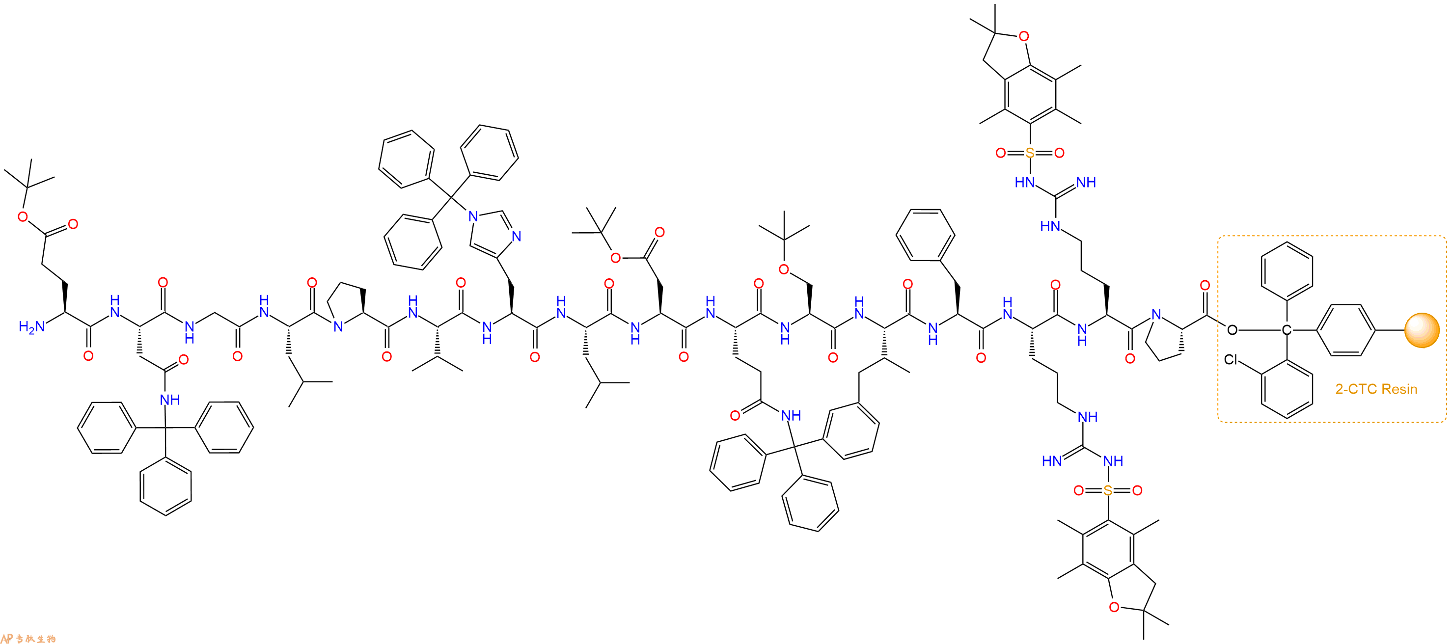
最后再经过步骤二得到 H2N-Glu(OtBu)-Asn(Trt)-Gly-Leu-Pro-Val-His(Trt)-Leu-Asp(OtBu)-Gln(Trt)-Ser(tBu)-Ile-Phe-Arg(Pbf)-Arg(Pbf)-Pro-CTC Resin,结构如下:
5、切割:6倍树脂体积的切割液(或每1g树脂加8ml左右的切割液),摇床摇晃 2小时,过滤掉树脂,用冰无水乙醚沉淀滤液,并用冰无水乙醚洗涤沉淀物3次,最后将沉淀物放真空干燥釜中,常温干燥24小试,得到粗品H2N-Glu-Asn-Gly-Leu-Pro-Val-His-Leu-Asp-Gln-Ser-Ile-Phe-Arg-Arg-Pro-COOH。结构图见产品结构图。
切割液选择:1)TFA:H2O=95%:5%
2)TFA:H2O:TIS=95%:2.5%:2.5%
3)三氟乙酸:茴香硫醚:1,2-乙二硫醇:苯酚:水=87.5%:5%:2.5%:2.5%:2.5%
(前两种适合没有容易氧化的氨基酸,例如Trp、Cys、Met。第三种适合几乎所有的序列。)
6、纯化冻干:使用液相色谱纯化,收集目标峰液体,进行冻干,获得蓬松的粉末状固体多肽。不过这时要取小样复测下纯度 是否目标纯度。
7、最后总结:
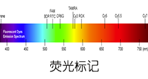
杭州专肽生物技术有限公司(ALLPEPTIDE https://www.allpeptide.com)主营定制多肽合成业务,提供各类长肽,短肽,环肽,提供各类修饰肽,如:荧光标记修饰(CY3、CY5、CY5.5、CY7、FAM、FITC、Rhodamine B、TAMRA等),功能基团修饰肽(叠氮、炔基、DBCO、DOTA、NOTA等),同位素标记肽(N15、C13),订书肽(Stapled Peptide),脂肪酸修饰肽(Pal、Myr、Ste),磷酸化修饰肽(P-Ser、P-Thr、P-Tyr),环肽(酰胺键环肽、一对或者多对二硫键环),生物素标记肽,PEG修饰肽,甲基化修饰肽等。
以上所有内容,为专肽生物原创内容,请勿发布到其他网站上。