浙江省科技型企业---加速您的多肽研究
首页 >多肽产品 >基质金属蛋白酶MMP FRET Substrate V、缓激肽MCA-(Ala7, Lys(DNP)9)-Bradykinin

多肽产品

323577-36-2,基质金属蛋白酶MMP FRET Substrate V、缓激肽Mca-(Ala⁷,Lys(Dnp)⁹,MCA-Arg-Pro-Pro-Gly-Phe-Ser-Ala-Phe-Lys(Dnp)-COOH,Mca-RPPGFSAF-K(Dnp)-OH,杭州专肽生物的产品

基质金属蛋白酶MMP FRET Substrate V、缓激肽MCA-(Ala7, Lys(DNP)9)-Bradykinin

内皮素转化酶-1(ECE-1)是一种非常敏感且内部淬灭的荧光底物,ECE-1是一种膜结合的锌金属肽酶,在氨基酸序列上与奈普利溶素有关。

编号:200072

CAS号:323577-36-2

单字母:Mca-RPPGFSAF-K(Dnp)-OH

纠错
  • 编号:200072
    中文名称:基质金属蛋白酶MMP FRET Substrate V、缓激肽MCA-(Ala7, Lys(DNP)9)-Bradykinin
    英文名:MCA-(Ala7, Lys(DNP)9)-Bradykinin
    CAS号:323577-36-2
    单字母:Mca-RPPGFSAF-K(Dnp)-OH
    三字母:MCA

    (7-甲氧基香豆素-4-基)乙酰基 MCA是一种荧光染料,其激发波长为325纳米,发射波长为392纳米。

    -Arg

    L-精氨酸:arginine。系统命名为(2S)-氨基-5-胍基戊酸。在生理条件下带正电荷,为编码氨基酸。是幼小哺乳动物的必需氨基酸。符号:R,Arg。

    -Pro

    L-脯氨酸:proline。系统命名为吡咯烷-(2S)-羧酸。为亚氨基酸。是编码氨基酸。在肽链中有特殊作用,如易形成顺式的肽键等。符号:P,Pro。

    -Pro

    L-脯氨酸:proline。系统命名为吡咯烷-(2S)-羧酸。为亚氨基酸。是编码氨基酸。在肽链中有特殊作用,如易形成顺式的肽键等。符号:P,Pro。

    -Gly

    甘氨酸:glycine。系统命名为 2-氨基乙酸。是编码氨基酸中没有旋光性的最简单的氨基酸,因具有甜味而得名。符号:G,Gly。

    -Phe

    L-苯丙氨酸:phenylalanine。系统命名为(2S)-氨基-3-苯基丙酸。是编码氨基酸。是哺乳动物的必需氨基酸。符号:F,Phe。

    -Ser

    L-丝氨酸:serine。系统命名为(2S)-氨基-3-羟基丙酸。是编码氨基酸。因可从蚕丝中获得而得名。符号:S,Ser。在丝原蛋白及某些抗菌素中含有 D-丝氨酸。

    -Ala

    丙氨酸:alanine。L-丙氨酸的系统命名为(2S)-氨基丙酸,是编码氨基酸,也叫L-α-丙氨酸。符号:A,Ala。D-丙氨酸存在于多种细菌细胞壁的糖肽中。β-丙氨酸是维生素泛酸和辅酶A的组分。

    -Phe

    L-苯丙氨酸:phenylalanine。系统命名为(2S)-氨基-3-苯基丙酸。是编码氨基酸。是哺乳动物的必需氨基酸。符号:F,Phe。

    -Lys(Dnp)

    侧链Dnp保护的赖氨酸

    -OH

    C端羧基:C-terminal carboxyl group。在肽或多肽链中含有游离羧基的氨基酸一端。在表示氨基酸序列时,通常将C端放在肽链的右边。

    氨基酸个数:9
    分子式:C66H81O19N15
    平均分子量:1388.44
    精确分子量:1387.58
    等电点(PI):-
    pH=7.0时的净电荷数:1
    平均亲水性:-0.44
    疏水性值:-0.19
    消光系数:-
    来源:人工化学合成,仅限科学研究使用,不得用于人体。
    储存条件:负80℃至负20℃
    标签:缓激肽(Bradykinin)    酶底物肽(Substrate Peptide)    侧链保护基肽    基质金属蛋白酶(MMP)    阿尔兹海默症(Alzheimer's Disease)    MCA标记肽   

  • 内皮素转化酶-1(ECE-1)是一种非常敏感且内部淬灭的荧光底物,ECE-1是一种膜结合的锌金属肽酶,在氨基酸序列上与奈普利溶素有关。ECE-1水解的kcal/Km值为1.9 x 10^7 M-1s-1,奈普利溶素水解的kcal/Km值为1.7 x 10^6 M-1s-1。对于MMP-2、MMP-9,kcat/Km值在10^4 M-1s-1的范围内,而对于MMP-1,没有观察到水解。该肽被基质分解素1(MMP-3)快速水解,kcat/Km=218000 M-1s-1,被明胶酶B(MMP-9)非常缓慢水解(kcat/Km=10100 M-1s-1)。未观察到MMP-1和MMP-2的水解。

    Very sensitive and internally quenched fluorescent substrate for endothelin-converting enzyme-1 (ECE-1), a membrane-bound zinc metallopeptidase that is related to neprilysin in amino acid sequence. The kcat/Km value for the hydrolysis by ECE-1 was 1.9 x 10^7 M-1s-1 and 1.7 x 10^6 M-1s-1 for the hydrolysis by neprilysin. For MMP-2, MMP-9 the kcat/Km values are in the range of 10^4 M-1s-1 whereas for MMP-1 there was no hydrolysis observed. This peptide was hydrolyzed rapidly by stromelysin 1 (MMP-3) with kcat/Km = 218000 M-1s-1 and very slowly by gelatinase B (MMP-9) (kcat/Km=10100 M-1s-1). There was no hydrolysis by MMP-1 and MMP-2 observed.

    Definition

    Bradykinin is a nonapeptide that is mainly found in animal preparations that are treated with the venom of the snake, Bothrops jararaca1,2.  It dialates blood vessels that in turn leads to decrease in blood pressure2. Bradykinin analogs are slightly modified structural derivatives of bradykinin that perform similar functions as bradykinin3.

    Discovery

    Bradykinin was discovered in the blood plasma of animals that were treated with the venom from the Brazilian snake, Bothrops jararaca1,2.  The discovery was part of a study that was related to toxicology of snake bites.  Bradykinin analogs were synthesized by solid-phase techniques in 1975 and their function was studied in rats and rabbits3.

    Classification

    Bradykinin is a 9 amino acid peptide that belongs to the kinin family of proteins4.  It has homologs in several animals including other snakes, frog, dog and humans4.

    Structural Characteristics

    Bradykinin has the sequence Arg-Pro-Pro-Gly-Phe-Ser-Pro-Phe-Arg3.  Several analogs of bradykinin have been synthesized. They are also nanopeptides containing substitutions of various amino acids of bradykinin. For example two analogs of bradykinin were synthesized one with 7-beta-homo-L-proline and the other with 8-beta-homo-L-phenylalanine substitutions3.  It was found that both of them are resistant to enzymatic degradation3.

    Mode of action

    Bradykinin binds to two different kinin G protein coupled receptors- B1 and B25.  Upon binding to these receptors it induces conversion of GTP to GDP which in turn triggers the conversion ATP to cAM which then acts as a second messenger resulting in the activation of genes.  B1 receptor is expressed as a result of tissue injury and is found to play a role in inflammation while B2 receptor participates in the vasodilatory role of bradykinin5,6.  Bradykinin analogs function is a similar fashion although depending on their structure they might have varying affinities to the receptors compared to bradykinin7. Also analogs of bradykinin have been synthesized that are specific to one of these receptors7.

    Functions

    Bradykinin is a potent endothelium-dependent vasodilator, causes contraction of non-vascular smooth muscle, increases vascular permeability and also is involved in the mechanism of pain. Bradykinin also causes natriuresis, contributing to a drop in blood pressure8. Bradykinin raises internal calcium levels in neocortical astrocytes causing them to release glutamate9. Overactivation of bradykinin is thought to play a role in a rare disease called Hereditary Angioedema, also known as Hereditary Angio-Neurotic Edema10. 

    Some analogs of bradykinin have been found to have prolonged hypotensive action compared to bradykinin (Eg: beta-H-Pro-bradykinin)3.  Some analogs have relative or even lower potencies compared to bradykinin (Eg: HArg1-Bradykinin and HArg9 Bradykinin)7.  Other analogs have been studied for their potential of finding bradykinin antagonists that might be useful in the treatment of angio-neurotic edema.  

    References

    1.     Partridge, SM (1948). (Title or abstract not available), Biochem. J., 42, 238.

    2.     Allen PK, Kusumam J, Yoji S, Yoshitaka N, Berhane G, Sesha R and Michael S (1998). Bradykinin formation: Plasma and tissue pathways and cellular interactions. Clinical reviews in allergy and immunology, 16, 4, 403-429.

    3.     Ondetti MA, Engel SL (1975). Bradykinin analogs containing beta.-homoamino acid, J. Med. Chem.,18 (7), 761–763.

    4.     Roseli A, Gomes S, Jair RC, Luis J and Valdemar H (1996). Met-Lys-Bradykinin-Ser, the kinin released from human kininogen by human pepsin. Immunopharmacology, 32, 76-79.

    5.    Peter GM, Amrita A, and Mauro P (2000). Association between Kinin B1 Receptor Expression and Leukocyte Trafficking across Mouse Mesenteric Postcapillary Venules. J Exp. Med., 192, 367-380.

    6.     Duchene J, Lecomte F, Ahmed S, Cayla C, Pesquero J, Bader M, Perretti M and Ahluwalia A, (2007). A Novel Inflammatory Pathway Involved in Leukocyte Recruitment: Role for the Kinin B1 Receptor and the Chemokine CXCL5. J Immunol., 179, 4849-4856.

    7.     Max ES, Phyllis AL (1974). Synthesis and pharmacology of homoarginine bradykinin analog. J. Med. Chem., 17 (11), pp 1227–1228.

    8.     Dendorfer A, Wolfrum S, Wagemann M, Qadri F, Dominiak P, (2001). Pathways of bradykinin degradation in blood and plasma of normotensive and hypertensive rats. Am J Physiol Heart Circ Physiol., 280:H2182

    9.     Kuoppala A, Lindstedt KA, Saarinen J, Kovanen PT, Kokkonen JO (2000). Inactivation of bradykinin by angiotensin-converting enzyme and by carboxypeptidase N in human plasma. Am J Physiol Heart Circ Physiol, 278(4):H1069-74.

    10.   Bas M, Adams V, Suvorava T, Niehues T, Hoffmann TK, Kojda G (2007). Nonallergic angioedema: role of bradykinin. Allergy, 62(8):842-56.

     Caspase酶对应的底物,Caspases(半胱氨酸天冬氨酸蛋白酶,半胱氨酸依赖性天冬氨酸定向蛋白酶)是一类蛋白酶家族,其功能与凋亡(程序性细胞死亡),坏死和发烧(炎症)的过程密切相关。

           什么是胱天蛋白酶?

          胱天蛋白酶(Caspases)是含半胱氨酸的天冬氨酸蛋白水解酶,它们是为细胞凋亡的主要介质。多种受体,例如TNF-α 受体,FasL受体,TLR和死亡受体,以及Bcl-2和凋亡抑制剂(IAP)蛋白家族参与并调节该caspase依赖性凋亡途径。一旦Caspase受到上游信号(外部或内在)刺激被激活,即会参与执行下游蛋白底物的水解作用,并触发一系列事件,导致细胞分解,死亡,吞噬作用和细胞碎片的清除。

          人Caspases酶

          人的Caspases家族基于序列相似性和生物学功能等共性主要可分为三大类:第一类由具有长胱天蛋白酶募集结构域的“炎症”胱天蛋白酶组成,他们对P4位上的较大的芳香族或疏水性残基具有亲和力。第二类由具有短的前体结构域的“细胞凋亡效应”胱天蛋白酶组成,而第三类由具有长的前提结构域的Pap位置具有亮氨酸或缬氨酸底物亲和力的“凋亡引发剂”胱天蛋白酶组成(表1)。

           表1. 人胱天蛋白酶的功能分类:

    细胞死亡途径 半胱天冬酶类型 酵素 物种
    细胞凋亡 启动器 Caspases 2 人与鼠
    细胞凋亡 启动器 Caspases 8 人与鼠
    细胞凋亡 启动器 Caspases 9 人与鼠
    细胞凋亡 启动器 Caspases 10 人的
    细胞凋亡 效应器 Caspases 3 人与鼠
    细胞凋亡 效应器 Caspases 6 人与鼠
    细胞凋亡 效应器 Caspases 6 人与鼠
    细胞焦亡 炎性的 Caspases 1 人与鼠
    细胞焦亡 炎性的 Caspases 4 人的
    细胞焦亡 炎性的 Caspases 5 人的

           启动器Caspase和效应器Caspase酶

          根据其在凋亡胱天蛋白酶途径中的作用,胱天蛋白酶可分为两类:启动器和效应器Caspase酶。启动器和效应器Caspas酶都具有由小亚基和大亚基组成的催化位点,Caspase酶的识别位

          凋亡启动器Caspase酶,例如caspase-2,-8,-9和-10可以启动caspase激活级联反应。Caspase-8对于形成死亡诱导信号复合物(DISC)是必不可少的,并且在激活后,Caspase-8激活下游效应子Caspase(例如Caspase 3)并介导线粒体中细胞色素c的释放。Caspase-8已被证明对IETD肽序列具有相对较高的底物选择性。凋亡效应胱天蛋白酶例如Caspase-3,-6和-7虽然不负责启动级联途径,但是当被激活时,它们在级联的中间和后续步骤中起着不可或缺的作用。Caspase-3(CPP32 / apopain)是关键效应器,因为它放大了来自启动器Caspase的信号,使用对Caspase-3有选择性的DEVD肽序列对活化的Caspase-3进行检测,可以检测Caspase-3的活性。

           Caspase酶底物和抑制剂

          Caspase底物和抑制剂由两个关键成分组成:Caspase识别序列和信号产生或蛋白酶抑制基序。不同Caspase识别序列不同,一般由三个或四个氨基酸组成(表2)。Caspase酶识别序列的N端通常有乙酰基(Ac)或碳苯甲氧基(Z)基团修饰,以增强膜的通透性。对应的Caspase识别特定的肽序列为其酶促反应切割位点,释放产生信号或抑制信号的基序。Caspase的显色和荧光底物均以相似的方式起作用,其中底物的信号或颜色强度与蛋白水解活性成正比。

           表2. Caspase的底物及其序列

    多肽 氨基酸序列 对应的Caspase的种类
    IETD Ile-Glu-Thr-Asp Caspase 8,颗粒酶B
    DEVD Asp-Glu-Val-Asp Caspase 3、6、7、8或10
    LEHD Leu-Glu-His-Asp Caspase 9
    VAD Val-Ala-Asp Caspase 1、2、3、6、8、9或10

             Caspase酶的显色底物

          Caspase的显色底物是有Caspase识别序列及生色基团组成,常见的生色团有pNA(对硝基苯胺或4-硝基苯胺),可使用酶标仪或分光光度计在405 nm处进行光密度检测。

           表3. Caspase的显色底物

    底物 Caspase 吸收(nm) 颜色
    Ac-DEVD-pNA * CAS 189950-66-1 * 半胱天冬酶3 405 nm 黄色
    Z-DEVD-pNA 半胱天冬酶3 405 nm 黄色
    Z-IETD-pNA * CAS 219138-21-3 * 半胱天冬酶8,颗粒酶B 405 nm 黄色

           Caspase的荧光底物

          Caspase的荧光底物的结构包含与半胱天冬酶识别相关的荧光团,例如7-氨基-4-甲基香豆素(AMC),7-氨基-4-三氟甲基香豆素(AFC), Rhodamine 110(R110)或ProRed™620。R110的Caspase底物比基于香豆素的Caspase底物(例如AMC和AFC)更敏感,但由于两步裂解过程,其动态范围更窄。 建议将R110标记的Caspase底物用于终点法测定,而将AMC和AFC标记的 Caspase底物用于动力学测定。

          图.从左到右,分别是AMC(7-氨基-4-甲基香豆素),AFC(7-氨基-4-三氟甲基香豆素),Rhodamine 110(R110)和ProRed™620的激发和发射光谱。

           表4.荧光半胱天冬酶底物。

    底物名称 对应的Caspase Ex(nm) Em(nm) ε¹ Φ²
    Ac-DEVD-AFC * CAS 201608-14-2 * 半胱天冬酶3、7 376 482 17000 0.53
    Ac-DEVD-AMC * CAS 169332-61-0 * 半胱天冬酶3、7 341 441 19000 N / D
    Z-DEVD-AFC 半胱天冬酶3、7 376 482 17000 0.53
    Z-DEVD-AMC * CAS 1135416-11-3 * 半胱天冬酶3、7 341 441 19000 N / D
    Z-DEVD-ProRed™620 半胱天冬酶3、7 532 619 N / D N / D
    (Z-DEVD)2 -R110 * CAS 223538-61-2 * 半胱天冬酶3、7 500 522 80000 N / D
    Z-DEVD-ProRed™620 半胱天冬酶3、7 532 619 N / D N / D
    Ac-IETD-AFC * CAS 211990-57-7 * 半胱天冬酶8,颗粒酶B 376 482 17000 0.53
    Z-IETD-AFC * CAS 219138-02-0 * 半胱天冬酶8,颗粒酶B 376 482 17000 0.53

           注意:

            1.ε=在其最大吸收波长处的摩尔消光系数(单位= cm -1-1)。

          2.Φ=水性缓冲液(pH 7.2)中的荧光量子产率。

           Caspase抑制剂

          Caspase抑制剂能与Caspase的活性位点结合并形成可逆或不可逆的连接,通常,Caspase抑制剂的结构由Caspase识别序列,诸如醛(-CHO)或氟甲基酮(-FMK)的官能团组成。具有醛官能团的胱天蛋白酶抑制剂是可逆的,而具有FMK的抑制剂是不可逆的。半胱天冬酶底物和抑制剂都具有较小的细胞毒性作用,因此,它们是研究半胱天冬酶活性的有用工具。

           表5. 可逆和不可逆的Caspase酶抑制剂

    抑制剂 Caspase的种类 是否可逆 Ex(nm) Em(nm)
    Ac-DEVD-CHO * CAS 169332-60-9 * 半胱天冬酶3、7 可逆的 -- --
    Ac-IETD-CHO * CAS 191338-86-0 * 半胱天冬酶8 可逆的 -- --
    mFluor™450-VAD-FMK 半胱天冬酶1,2,3,6,8,9,10 不可逆的 406 445
    mFluor™510-VAD-FMK 半胱天冬酶1,2,3,6,8,9,10 不可逆的 412 505
    FITC-C6-DEVD-FMK 半胱天冬酶3、7 不可逆的 491 516
    FITC-C6-DEVD-FMK 半胱天冬酶3、7 不可逆的 491 516
    FITC-C6-LEHD-FMK 半胱天冬酶9 不可逆的 491 516
    FITC-C6-LEHD-FMK 半胱天冬酶9 不可逆的 491 516
    FAM-VAD-FMK 半胱天冬酶1,2,3,6,8,9,10 不可逆的 493 517
    SRB-VAD-FMK [磺胺丁胺B-VAD-FMK] 半胱天冬酶1,2,3,6,8,9,10 不可逆的 559 577

    定义

    基质金属蛋白酶(MMP)属于锌内肽酶家族,统称为metzincins。metzincin超家族的特征是高度保守的基序,该基序包含在催化位点与锌结合的三个组氨酸和位于活性位点下方的保守的蛋氨酸。

    发现

    间质胶原酶,首先被确定的MMP家族成员,最初是在旨在解释designed变态为青蛙1的胶原重塑的实验中发现的。MMP家族由20多个共享相同功能域的相关锌依赖性酶组成。这些酶既具有通常基于优选底物的描述性名称,又具有基于发现顺序的MMP编号系统1。

    结构特征

    MMP的基本结构由以下同源结构域组成:1)将MMP引导至分泌或质膜插入途径的信号肽;2)前结构域,通过占据活性位点锌使酶具有潜伏期,使底物难以接近催化酶;3)含锌的催化域;4)溶血毒素域,介导与底物的相互作用并赋予酶特异性;5)连接催化和血红蛋白结构域的铰链区。MMP7或基质溶素是最小的MMP,缺少血红素结构域,但在底物降解中表现出特异性。额外的结构域和底物特异性导致MMP分为亚组。

    MMP的蛋白质结构中两个序列基序高度保守。在所有MMP的催化域中发现的共有基序HExGHxxGxxH,包含3个与活性中心的锌离子(Zn)配位的组氨酸。PRCGxPD基序位于MMPs前结构域的C末端;该基因座的半胱氨酸残基(C)与活性中心的锌原子的配位赋予了酶2、3潜伏期。

    行动方式

    MMP的体内活性在几个水平上受到严格控制。这些酶通常以极低的量表达,其转录受到细胞因子和生长因子(如白介素(IL-1,IL-4,IL-6),转化生长因子(EGF,HGF,TGFβ)的正向或负向严格调控。 ),或肿瘤坏死因子α(TNFα的)4, 5。这些调节分子中的一些可以被MMP蛋白水解激活或失活(反馈作用)。转录后,MMP活性受到位于新合成的酶的N末端的前肽所赋予的潜伏期的限制。从细胞分泌后,MMPs的激活取决于前结构域与催化位点的相互作用的破坏,这可能通过前结构域的构象变化或蛋白水解去除而发生。在其前肽中含有弗林蛋白酶样识别结构域的MMP(MMP11,MT-MMP,MMP28)可以通过枯草杆菌蛋白酶丝氨酸蛋白酶家族的成员在反式高尔基体网络中被激活。MMP14在细胞表面的proMMP2激活中起着不可或缺的作用。分泌的MMP的胞外蛋白水解激活可以通过丝氨酸蛋白酶(如纤溶酶)介导,这暗示着这两个酶基团在ECM重塑中具有相互依赖性。某些活动的MMP可以激活其他proMMP,例如MMP9和MMP1的MMP3激活。激活后,MMP会受到内源性抑制剂,自降解和选择性内吞作用的进一步调节。已经证明通过低密度脂蛋白受体相关蛋白(LRP)机制内吞MMP2、9和137。

    功能

    MMP参与癌症的各个方面-MMP诱导的肝素结合上皮生长因子,胰岛素样生长因子和成纤维细胞生长因子从细胞表面释放(脱落),促进细胞增殖。另一方面,由MMP释放和激活ECM螯合的TGFβ可以导致细胞增殖受到抑制。

    炎症性疾病-许多报道暗示MMP1,MMP3和MMP9参与类风湿和骨关节炎。

    心血管疾病-大量研究表明,动脉粥样硬化和动脉瘤形成部位6的MMP,尤其是MMP9水平升高。已经提出MMP代表冠状动脉疾病患者中炎症的敏感标志。

    肺部疾病- MMP的水平升高已经牵涉在各种肺部疾病,包括急性呼吸窘迫综合征,哮喘,支气管扩张和囊性纤维化的病理生理学。MMP,EMMPRIN和TIMPs是由肺中的许多常驻细胞产生的,因此使它们在疾病中的作用7的分析变得复杂。

    中枢神经系统疾病-在观察到MMP9在类似于多发性硬化症和格林-巴利综合症的动物模型中的关键作用之后,MMPs参与了几种不同类型的神经系统疾病。

    休克综合征- MMP8和MMP9存储在多形核白细胞的颗粒。这些细胞是炎症和感染过程中的关键效应器。这些MMP在休克中的作用得到了对MMP9缺陷型小鼠的研究的支持,这些小鼠被证明对内毒素休克具有抗性。Dubois等人[ 8]提出,特定的MMP9抑制作用是治疗败血性休克综合症的一种潜在方法。 

    参考

    1.     Gross J, Lapiere CM (1962). Collagenolytic activity in amphibian tissues; a tissue culture assay. PNAS., 48:1014-1022.

    2.     Nagase H, Woessner F (1999). Matrix metalloproteinases. J Biol Chem., 274(31):21491-21494.

    3.     Birkedal-Hansen H. (1995). Proteolytic remodeling of extracellular matrix. Curr Opin Cell Biol., 7:728-735.

    4.     Zucker S, Pei D, Cao J, Lopez-Otin C (2003). Membrane type-matrix metalloproteinases (MT-MMP). Cell Surface Proteases., 54:1-74.

    5.     Yang Z, Strickland DK, Bornstein P (2001). Extracellular MMP-2 levels  are regulated by the low-density lipoprotein-related scavenger receptor and thrombospondin. J Biol Chem., 276: 8403-8408.

    6.     Van den Steen PE, Dubois B, Nelissen I, Rudd PM, Dwek RA, Opdenakker G (2002). Biochemistry and molecular biology of gelatinase B or matrix metalloproteinase-9 (MMP-9). Crit Rev Biochem Mol Biol,. 37:376-536.

    7.     Haseneen N, Vaday G, Zucker S, Foda HD (2003). Mechanical stretch induces MMP-2 release and activation in lung endothelium: role of EMMPRIN. Am J Physio Lung Cell Mol Physiol., 165:541-547.

    8.     Dubois B, Starckx S, Pagenstecher A, Oord J, Arnold B, Opdenakker G. Gelatinase B (2002). Deficiency protects against endotoxin shock. Eur J Immunol., 32:2163-2171.

    Extracellular amyloid-β peptide deposition into cerebellar plaques and formation of intracellular neurofibrillary fibers accompanied by the loss of neurons are characteristic histopathological lesions found in the brains of Alzheimer‘s disease patients. Individuals suffering from this disease show a gradual loss of cognitive functions and disturbances in behavior. Apart from some rare familial forms of the disease, the onset of Alzheimer‘s disease is usually above 60 years. Since the risk to develop the disease increases with age, Alzheimer‘s disease has turned into a major health and social problem in “first world” countries with an increasing proportion of older people, and is going to become one in emerging states. In this brochure we present amyloid peptides and related products for Alzheimer‘s disease research.

    ALZHEIMER’S DISEASE
    Alzheimer‘s disease (AD) is the prevalent cause of dementia in elderly people and has become one of the leading causes of death in developed countries together with cardiovascular disorders, cancer, and stroke. It is estimated that more than 46 millions of people suffer from AD all over the world. As age advances, the risk for developing AD increases. The frequency of AD at the age of 60-64 is about 1% and doubles approximately every five years. By the age of 90 and older, approximately 50% of the population suffers from this disease. AD is an irreversible and progressive neurodegenerative disorder. Symptoms include gradual loss of cognitive functions such as memory, verbal and visuospatial abilities, changes in personality, behavior, and activities of daily living. AD patients in the final stages are completely dependent on the care of others.

    The characteristic lesions in the brains of AD patients were first described by the German neuropsychiatrist Alois Alzheimer in 1906 during the post-mortem examination of a mentally ill patient whose deterioration he had observed until her death. The lesions consisted of dense extracellular deposits, now designated as neuritic or senile plaques, and intracellular dense bundles of fibrils, which are now known as neurofibrillary tangles.

    Currently, diagnosis of AD with adequate testing is approximately 90% accurate. It is based on the exclusion of a variety of diseases causing similar symptoms and a careful neurological and psychiatric examination, as well as neuropsychological testing. Imaging technologies for detecting amyloid plaques and tangles in vivo are becoming more precise and thus a valuable additional tool. Numerous potential biomarkers as α1 -antitrypsin, complement factor H, α2 -macroglobulin, apolipoprotein J, and apolipoprotein A-I for diagnosing AD are being evaluated. However, post-mortem histopathological examination of the brain is still the only definite diagnosis of this disease.

    AD can be either inherited or sporadic. The inherited or familial AD is rare and comprises only 5-10% of all cases. Autosomal dominant mutations in the amyloid β/A4 protein precursor (APP) gene on chromosome 21 and the presenilin-1 or -2 genes on chromosomes 14 and 1, respectively, have been attributed to the early onset (before the age of 65) of this disease.

    APP belongs to the type-1 integral membrane glycoproteins with at least 10 isoforms generated by alternative splicing of the 19 exons. The predominant transcripts are APP695, APP751, and APP770. A number of mutations within the APP gene have been detected in families with an inherited risk for early onset of AD. Usually, they are named after the region, in which they have been detected, e.g. the London APP717 mutations (V717I, V717F, V717G), the Swedish APP670/671 double mutation (K670N/M671L), the Flemish APP692 mutation (A692G), or the Dutch APP693 mutation (E693Q). The Swedish mutation of the β-secretase cleavage site of APP and mutations of positions 692-694 (Aβ 21-23), which strongly influence the aggregation behavior of Aβ, have been studied intensively.

    A choice of relevant mutations in the Aβ region of APP is assembled in the table below.

    Exchanged Position in APP Exchanged Position in Aβ Designation
    A673T A2T Icelandic
    H677R H6R English
    D678H D7H Taiwanese
    D678N D7N Tottori
    A692G A21G Flemish
    E693D E22∆ Osaka
    E693G E22G Arctic
    E693Q E22Q Dutch
    E693K E22K Italian
    D694N D23N Iowa
    L705V L34V Piedmont


    The presenilins are another group of proteins involved in the development of AD. Presenilins are integral membrane proteins with eight transmembrane domains localized in the endoplasmic reticulum and the Golgi apparatus. A multitude of mutations within the presenilin-1 and two within the presenilin-2 gene account for most of the cases of early onset of AD.

    Genetic factors may contribute as well to the late onset of AD. Increased susceptibility is associated with the expression of different apolipoprotein E (ApoE) isoforms due to the polymorphism in the APOE gene on chromosome 19. In the central nervous system, ApoE has been implicated in growth and repair during development or after injury. Carriers of the APOEε4 allele show a higher risk in developing the disease than carriers of the other two possible alleles APOEε2 and APOEε3. The ApoEε4 effect seems to be dose-dependent since individuals with two of these alleles seem to be at two-fold higher risk to develop the disease than those with one allele. Polymorphisms of the α2 -macroglobulin gene on chromosome 12 and the gene coding low-density lipoprotein receptor-related protein 1 (LRP1), LRP1-C/T, have also been suggested to be a risk factor for the late onset of AD. However, further studies in this field are required.

    A number of additional, most diverse risk factors have been proposed. These include gender, ethnic group, head trauma, cardiovascular diseases, and educational level.

    AD THERAPEUTIC STRATEGIES RELY ON DETAILED KNOWLEDGE OF THE MOLECULES INVOLVED

    Women, Hispanics, individuals who have experienced a head trauma earlier in life, and persons who suffer from cardiovascular diseases appear to have a higher risk of developing the disease.

    The etiology of AD is still not completely understood. Initial research focused upon determining the molecular structure of the senile plaques and the neurofibrillary tangles originally described by Alois Alzheimer. The main constituents of the senile plaques were identified as cleavage products of APP, designated as amyloid β-peptides (Aβ peptides).

    Depending on the composition and the fraction of fibrillar to non-fibrillar forms of these amyloid peptides, several kinds of senile plaques can be distinguished. Three types of proteases, α-secretase, β-secretase (or β-site APP-cleaving enzyme, BACE), and γ-secretase are involved in APP processing. APP can either be processed by the α- and γ- or by the β- and γ-secretases. The major two amyloid peptides identified in senile plaques, amyloid β-protein (1-40) (Aβ40) and amyloid β-protein (1-42) (Aβ42), are generated by successive proteolysis of APP by β- and γ-secretases. Cleavage of APP by β-secretase results in the release of the extracellular N-terminal protein fragment known as soluble APP-β molecule (sAPP-β). Then, the membrane-retained APP is further processed within the transmembrane domain by γ-secretase to yield either Aβ40 or Aβ42. The formation of Aβ40 and Aβ42 is a normal process, and both peptides can be detected in the plasma and cerebrospinal fluid (CSF) of healthy subjects.

    In most studies, similar concentrations of Aβ40 have been measured in the CSF of both healthy controls and AD patients. On the other hand, Aβ42 concentrations in the CSF of AD patients are significantly lower than in normal controls, probably reflecting an increased deposition as insoluble plaques.

    The neurofibrillary tangles found inside neurons of Alzheimer’s brains are composed of paired helical filaments whose main components are hyperphosphorylated forms of tau, a microtubule associated protein involved in promoting microtubule assembly and stabilization. Self-assembly into paired helical filaments is believed to be a result of hyperphosphorylation due to either the increased activity of protein kinases or the decreased activity of phosphatases.

    Several lines of evidence support the view that the accumulation of Aβ42 in the brain is a primary event in the development of AD. Increased cerebral Aβ production appears to be characteristic for all the mutations within the APP and the presenilin genes of familial AD. In patients with Down syndrome (trisomy 21), elevated levels of APP and Aβ due to a third copy of the APP gene result in deposition of Aβ at an early age between 20 and 30.

    Formation of neurofibrillary tangles is considered as a consequence of Aβ deposition with a further impact on the progression of the disease possibly due to disruption of axonal transport mechanisms in neurons.

    The detailed knowledge about the molecules involved in AD has led to the development of several therapeutic strategies.

    One strategy aims at the reduction of Aβ40 and Aβ42 by inhibition of either β- or γ-secretase activity or by clearance of Aβ in the brain by means of immunization with these peptides. Transition metals as Cu, Fe and Zn play an important role in the pathology of AD. Aggregation and neurotoxicity of Aβ are dependent on the presence of copper, so Cu-chelating agents showed promising effects in animal models. Another approach is the prevention of the cellular inflammatory response in the cerebral cortex elicited by the progressive accumulation of Aβ. Further preventive therapeutic strategies are based on the findings that cholesterol-lowering drugs such as statins and estrogen replacement therapy reduce the risk of developing AD. An additional treatment alternative would be the inhibition of the serine-threonine protein kinases, glycogen synthase kinase 3 (GSK3) and cyclin-dependent kinase 5 (CDK5), which are probably responsible for the phosphorylation of the tau protein. Inhibition of calpain, an enzyme showing increased activity in AD brains, led to promising results in animal studies. Calpain cleaves the CDK5 activator p35 leading to p25 formation and CDK5 overactivation.

    Several acetylcholinesterase inhibitors such as tacrine, donepezil, rivastigmine, and galantamine have been approved for the treatment of mild to moderate AD by the FDA and other authorities. They act by reducing the deficits of the neurotransmitter acetylcholine associated with cognitive impairment in AD patients. The amantadine derivative memantine, an NMDA receptor antagonist, which was already used for the treatment of moderate to severe AD in Europe, has gained approval in the United States by the FDA as well.

    A promising drug candidate, the β-secretase inhibitor verubecestat (MK-8931) developed for the management of mild to moderate AD, has moved to phase III. Moreover, the BACE inhibitor AZD3293 showed encouraging results in clinical studies. Antibodies as aducanumab and solanezumab, which have been designed to degrade plaques and lower the level of Aβ in the brain, have reached advanced stages of clinical testing for mild cases of AD.

    Despite the many promising therapeutic approaches, AD still remains a major burden for the patients, their relatives, and the society.

    MCA标记肽的说明

    (7-Methoxycoumarin-4-yl)acetyl is fluorophor with an excitation at 325 nm  and emission of 392 nm .

    MCA标记肽相关文献:

    Characterization of the Altai Maral Chymosin Gene, Production of a Chymosin Recombinant Analog in the Prokaryotic Expression System, and Analysis of Its Several Biochemical Properties.
    Belenkaya, S. V., A. A. Bondar, T. A. Kurgina, V. V. Elchaninov, A. Yu Bakulina, E. A. Rukhlova, O. I. Lavrik, A. A. Ilyichev, and D. N. Shcherbakov. Biochemistry (Moscow), 2020.

    IL-1b is an innate immune sensor of microbial proteolysis.
    LaRock, Christopher N., et al. Science Immunology 100.200: 300 (2016).

  • DOI名称
    10.1167/iovs.03-0769Characterization of a bradykinin-hydrolyzing protease from the bovine lens下载
    10.1042/BJ20041481Flexibility in substrate recognition by thimet oligopeptidase as revealed by denaturation studies下载
    10.1021/bi051035kTropolysin, a new oligopeptidase from African trypanosomes下载
    10.1161/01.HYP.0000237862.94083.45Enalapril attenuates downregulation of Angiotensin-converting enzyme 2 in the late phase of ventricular dysfunction in myocardial infarcted rat下载
    10.1038/nature05143Structures of human insulin-degrading enzyme reveal a new substrate recognition mechanism下载
    10.1016/j.jneumeth.2007.12.003Immunocapture-based fluorometric assay for the measurement of insulin-degrading enzyme activity in brain tissue homogenates下载
    10.1111/j.1742-4658.2008.06685.xHydrogen bond residue positioning in the 599-611 loop of thimet oligopeptidase is required for substrate selection下载
  • 多肽MCA-Arg-Pro-Pro-Gly-Phe-Ser-Ala-Phe-Lys(Dnp)-COOH的合成步骤:

    1、合成CTC树脂:称取1.21g CTC Resin(如初始取代度约为0.69mmol/g)和1.0mmol Fmoc-Lys(Dnp)-OH于反应器中,加入适量DCM溶解氨基酸(需要注意,此时CTC树脂体积会增大好几倍,避免DCM溶液过少),再加入2.5mmol DIPEA(Mw:129.1,d:0.740g/ml),反应2-3小时后,可不抽滤溶液,直接加入1ml的HPLC级甲醇,封端半小时。依次用DMF洗涤2次,甲醇洗涤1次,DCM洗涤一次,甲醇洗涤一次,DCM洗涤一次,DMF洗涤2次(这里使用甲醇和DCM交替洗涤,是为了更好地去除其他溶质,有利于后续反应)。得到  Fmoc-Lys(Dnp)-CTC Resin。结构图如下:

    2、脱Fmoc:加3倍树脂体积的20%Pip/DMF溶液,鼓氮气30分钟,然后2倍树脂体积的DMF 洗涤5次。得到 H2N-Lys(Dnp)-CTC Resin 。(此步骤脱除Fmoc基团,茚三酮检测为蓝色,Pip为哌啶)。结构图如下:

    3、缩合:取2.5mmol Fmoc-Phe-OH 氨基酸,加入到上述树脂里,加适当DMF溶解氨基酸,再依次加入5.01mmol DIPEA,2.38mmol HBTU。反应30分钟后,取小样洗涤,茚三酮检测为无色。用2倍树脂体积的DMF 洗涤3次树脂。(洗涤树脂,去掉残留溶剂,为下一步反应做准备)。得到Fmoc-Phe-Lys(Dnp)-CTC Resin。氨基酸:DIPEA:HBTU:树脂=3:6:2.85:1(摩尔比)。结构图如下:

    4、依次循环步骤二、步骤三,依次得到

    H2N-Phe-Lys(Dnp)-CTC Resin

    Fmoc-Ala-Phe-Lys(Dnp)-CTC Resin

    H2N-Ala-Phe-Lys(Dnp)-CTC Resin

    Fmoc-Ser(tBu)-Ala-Phe-Lys(Dnp)-CTC Resin

    H2N-Ser(tBu)-Ala-Phe-Lys(Dnp)-CTC Resin

    Fmoc-Phe-Ser(tBu)-Ala-Phe-Lys(Dnp)-CTC Resin

    H2N-Phe-Ser(tBu)-Ala-Phe-Lys(Dnp)-CTC Resin

    Fmoc-Gly-Phe-Ser(tBu)-Ala-Phe-Lys(Dnp)-CTC Resin

    H2N-Gly-Phe-Ser(tBu)-Ala-Phe-Lys(Dnp)-CTC Resin

    Fmoc-Pro-Gly-Phe-Ser(tBu)-Ala-Phe-Lys(Dnp)-CTC Resin

    H2N-Pro-Gly-Phe-Ser(tBu)-Ala-Phe-Lys(Dnp)-CTC Resin

    Fmoc-Pro-Pro-Gly-Phe-Ser(tBu)-Ala-Phe-Lys(Dnp)-CTC Resin

    H2N-Pro-Pro-Gly-Phe-Ser(tBu)-Ala-Phe-Lys(Dnp)-CTC Resin

    Fmoc-Arg(Pbf)-Pro-Pro-Gly-Phe-Ser(tBu)-Ala-Phe-Lys(Dnp)-CTC Resin

    以上中间结构,均可在专肽生物多肽计算器-多肽结构计算器中,一键画出。

    最后再经过步骤二得到 H2N-Arg(Pbf)-Pro-Pro-Gly-Phe-Ser(tBu)-Ala-Phe-Lys(Dnp)-CTC Resin,结构如下:

    5、7-甲氧基香豆素-4-乙酸(MCA)反应连接:在上述树脂中,加入适当DMF后,再加入2.5mmol 7-甲氧基香豆素-4-乙酸(MCA)到树脂中,再加入5.01mmol DIPEA、2.38mmol HBTU,鼓氮气反应30分钟。用2倍树脂体积的DMF 洗涤3次树脂(洗涤树脂,去掉残留溶剂,为下一步反应做准备)。 得到MCA-Arg(Pbf)-Pro-Pro-Gly-Phe-Ser(tBu)-Ala-Phe-Lys(Dnp)-CTCResin。 结构如下:

    6、切割:6倍树脂体积的切割液(或每1g树脂加8ml左右的切割液),摇床摇晃 2小时,过滤掉树脂,用冰无水乙醚沉淀滤液,并用冰无水乙醚洗涤沉淀物3次,最后将沉淀物放真空干燥釜中,常温干燥24小试,得到粗品MCA-Arg-Pro-Pro-Gly-Phe-Ser-Ala-Phe-Lys(Dnp)-COOH。结构图见产品结构图。

    切割液选择:1)TFA:H2O=95%:5%

    2)TFA:H2O:TIS=95%:2.5%:2.5%

    3)三氟乙酸:茴香硫醚:1,2-乙二硫醇:苯酚:水=87.5%:5%:2.5%:2.5%:2.5%

    (前两种适合没有容易氧化的氨基酸,例如Trp、Cys、Met。第三种适合几乎所有的序列。)

    6、纯化冻干:使用液相色谱纯化,收集目标峰液体,进行冻干,获得蓬松的粉末状固体多肽。不过这时要取小样复测下纯度 是否目标纯度。

    7、最后总结:

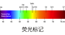
    杭州专肽生物技术有限公司(ALLPEPTIDE https://www.allpeptide.com)主营定制多肽合成业务,提供各类长肽,短肽,环肽,提供各类修饰肽,如:荧光标记修饰(CY3、CY5、CY5.5、CY7、FAM、FITC、Rhodamine B、TAMRA等),功能基团修饰肽(叠氮、炔基、DBCO、DOTA、NOTA等),同位素标记肽(N15、C13),订书肽(Stapled Peptide),脂肪酸修饰肽(Pal、Myr、Ste),磷酸化修饰肽(P-Ser、P-Thr、P-Tyr),环肽(酰胺键环肽、一对或者多对二硫键环),生物素标记肽,PEG修饰肽,甲基化修饰肽

    以上所有内容,为专肽生物原创内容,请勿发布到其他网站上。

  • 暂时没有数据